Electromyography
DEFINITION
Electromyography (EMG) is a diagnostic process used to determine the health of muscles, the nerve cells that control these muscles, or motor neurons.
In purpose, the EMG is an electrical recording of muscle activity that aids in diagnosing neuromuscular disease.
The Electromyography scan, sometimes also called a myogram, is used to identify the electrical signals from these motor neurons (which cause the muscles to move).
These electrical signals identified by the EMG allow the medical specialist to interpret such signals to diagnose any problems within the muscles and the nerves that control them.
DESCRIPTION
In the time of an EMG test, a delicate needle is injected into the muscle being tested.
This can cause some displeasure, similar to the sensation of receiving an injection.
Reportings are done while the muscle is resting and then along with the contraction duration.
The technician performing the testing can maneuver the limb being tested and instruct the patient to move it with variable levels of force.
The needle may be moved in the same muscle for further recording. Other muscles may be tested as well.
A bit different test, known as the nerve conduction velocity testing, is often done simultaneously and with unaltered equipment.
This test uses stimulating and reporting electrodes; short electrical shocks are administered to measure the capacity of the nerve to pass on electrical signals.
The test may invoke mild tingling and distress familiar to a soft shock, such as static electricity.
Induced potentials may also be performed for further diagnostic facts.
Nerve conducting velocity and evoked potential testing is beneficial when pain or sensory complaints are more prominent than weakness.
The EMG procedure will usually take 30–60 minutes to complete.
PURPOSE
Muscles are excited by signals traveling from nerve cells known as motor neurons.
This incentive causes electrical activity in the muscle, which in turn causes contraction. An electrode, which consists of a tiny, solid needle (pin), is inserted through the skin and muscle.
It is connected to a recording device that detects this electrical activity. When a needle is used, the process is called a needle EMG, or simply an EMG.
The needle electrode and recorder pair is called an electromyography machine, which includes a monitor called an oscilloscope.
A speaker is included, which provides crackling sounds as the electrical intensity rises and falls.
EMG can predispose whether a particular muscle responds appropriately to stimulation and whether a muscle remains inactive when not stimulated.
Thus, during the test, the patient will be asked to contract particular muscles, such as those in the leg.
The electrical wave produced on the EMG machine will determine the condition of the muscle and nerves as it responds to the contraction.
Usually, a nerve conduction velocity (NCV) test (or nerve conduction study) is also performed simultaneously as an Electromyography.
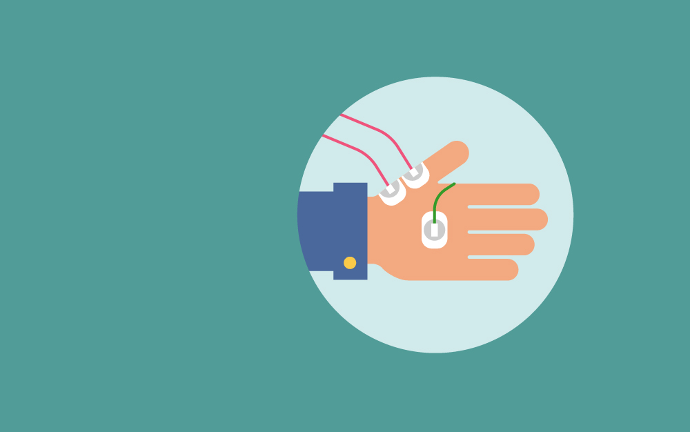
This test consists of two electrodes being placed apart on the surface of the skin. One electrode is activated so that a muscle is electronically simulated.
The second electrode senses the activity (electrical impulse) of the muscle as it moves.
The NCV test measures the intensity and speed at which electrical signals pass between two points.
The EMG procedure is implemented frequently to aid diagnose different illnesses causing weakness.
Albeit Electromyography is a procedure of testing the motor system, it can help pinpoint irregularities of nerves or spinal nerve roots, which may be connected with pain or insensibility.
EMG’s other symptoms can be useful include numbness, tingling, atrophy, stiffness, fasciculation (twitch), pain or cramping, deformity, and spasticity (abnormal muscle performance, such as muscle weakness).
Electromyography results may help decide whether symptoms are thanx to a muscle illness or a neurological disease and, once combined with clinical results, usually give a confident diagnosis.
EMG may help diagnose many muscles and nerve disorders, including (but not restricted to) the following:
- muscular dystrophy or polymyositis
- congenital myopathies
- myasthenia gravis
- mitochondrial myopathies
- metabolic myopathies
- myotonias
- peripheral neuropathies
- radiculopathies
- nerve lesions
- amyotrophic lateral sclerosis
- polio
- spinal muscular atrophy
- Guillain-Barré syndrome
- ataxias
- myasthenias
Only a few individual precautions are required for this test. Subjects with a history of bleeding disorder should consult with their treating physician before the test.
Any person taking blood-thinning medications should inform the neurologist or other medical professional conducting the EMG procedure.
An individual with a pacemaker or other electrical medical device should inform the medical team of this before the procedure.
If a muscle biopsy is outlined as part of the diagnostic work-up, the EMG should not be done at the same site, as it may affect the appearance of the muscle.
PREPARATION
Very few special preparations are needed before the EMG. Natural oils on the skin should be removed before the test.
Therefore, take a shower or bath before the procedure. In addition, do not apply creams or lotions.
The doctor overseeing and interpreting the test results should be given information regarding the symptoms, medical conditions, suspected diagnosis, neuroimaging studies, and other test results.
AFTERCARE
Minor bruising may occur after the procedure; it will fade over the next few days. Slight pain and bleeding may follow for a couple of hours after the test is completed.
The muscle can be tender for a day or two. If any of these do not go away after several days, contact a medical professional.
The doctor will not restrict activities after the test is completed under normal circumstances.
RESULTS
The results of the test are available instantly upon completion of the EMG. However, a trained medical specialist, such as a neurologist, must analyze and interpret the results.
Normal results
There may be certain short EMG activity at the moment of needle insertion.
This activity can be increased in illnesses of the nerve and declined in long-standing muscle deteriorations when muscle tissue is replaced by fibrous tissue or fat.
Muscle tissue normally does not show EMG activity when resting or when moved passively by the power of the examiner.
Once the patient actively contracts the muscle, spikes (motor unit action potentials) should appear on the recording screen, reflecting the electrical activity.
When the muscle is contracted more forcefully, more muscle fibers are recruited or activated, causing more EMG activity.
Abnormal results
The analysis of EMG results is not a plain matter, demanding analysis of the appearance, duration, amplitude, and other traits of the spike patterns.
RISKS
There are no significant risks in performing this procedure. The only minor risks occur when a needle is inserted under the skin.
Such risks may include pain or discomfort, bleeding, bruising, or infection.
Since medicine is not being injected under the skin, less pain is usually the case when compared to the insertion of a regular needle.
In addition, there is a minute risk for nerve injury when the needle is inserted under the skin.
When such an electrode is inserted into the chest wall, a small risk is present that the puncture could cause air to leak into the cavity between the lungs and the chest wall.
In such a case, a collapsed lung could result, although it is doubtful.
When the nerve conduction velocity test is performed, the patient will perceive a brief and very mild shock or tingling sensation.
Electrical activity at resting is abnormal; the certain firing template can hint at denervation (for instance, radiculopathy, a nerve lesion, or lower motor neuron degeneration), inflammatory myopathy, or myotonia.
Drops in the amplitude and extent of spikes are connected with muscle diseases, which also show quicker recruitment of other muscle fibers to compensate for weakness. Recruitment is reduced in nerve disorders.
KEY TERMS
- Motor neurons— Nerve cells that transmit signals from the brain or spinal cord to the muscles.
- Motor unit action potentials— Spikes of electrical activity recorded during an EMG reflect the number of motor units (motor neurons and the muscle fibers they transmit signals to) activated when the patient voluntarily contracts a muscle.
Resources
Daube, Jasper R., and Devon I. Rubin, eds. Clinical Neurophysiology. New York: Oxford University Press, 2009.
Kamen, Gary, and David A. Gabriel. Essentials of Electromyography. Champaign, IL: Human Kinetics, 2010.
Pease, William S., Henry L. Lew, and Ernest W. Johnson. Johnson’s Practical Electromyography. Philadelphia: Lip pincott Williams and Wilkins, 2007.
Emedicinehealth.com , WebMD. “Electromyography (EMG).” Last updated November 30, 2011. http://www.emedicinehealth.com/electromyography_emg/article_em.htm. (accessed October 14, 2014).
Mayo Clinic. “Electromyography (EMG).” Last updated October 25, 2012. http://www.mayoclinic.com/health/emg/MY00107. (accessed October 14, 2014).
Medline Plus, National Library of Medicine, and National Institutes of Health. “Electromyography.” (September 22, 2008), http://www.nlm.nih.gov/medlineplus/ency/article/003929.htm . (accessed October 14, 2014).
American Association of Neuromuscular and Electrodiagnostic Medicine, 2621 Superior Drive NW, Rochester, MN 55901, (507) 288-0100, (507) 288-1225 aanem@aanem.org, http://www.aanem.org/.
Richard Robinson


